Volume: 16 Issue: 1 February 2018
FULL TEXT
Objectives: Fluid therapy is required to maintain perfusion to donor organs. Recent reviews on the choices of fluids have emphasized the safety of using crystalloids, as opposed to fluid therapy with colloids, which has been reported to be either unequivocally or potentially harmful in a number of studies on various patient populations. We aimed to analyze whether the type of fluid administered to donors is connected with kidney transplant outcomes.
Materials and Methods: A total of 100 consecutive brain-dead multiorgan donors and their respective 181 kidney recipients were studied retrospectively. Data concerning donor fluid therapy, the charac-teristics of the donors and the recipients, and outcomes after kidney transplant were extracted from organ retrieval and patient records. Cases with early graft function were compared with cases with delayed graft function.
Results: Donors had received both crystalloids and colloids in most cases (84%). Fluid therapy with crystalloids alone was more common among the 40 recipients with delayed (30%) than in the 103 recipients with early graft function (11%) (P = .005). Donor age, time on renal replacement therapy before transplant, and donor fluid therapy with crystalloids alone were independent risk factors for delayed graft function in multivariate analysis.
Conclusions: Our results suggest that donor fluid therapy including colloids could be beneficial instead of harmful compared with treatment with crystalloids alone. This finding needs to be evaluated in pro-spective studies.
Key words : Delayed graft function, Fluid therapy, Renal transplant
Introduction
Every year, about 170 kidney transplant procedures are performed in Finland, with recipients most commonly requiring transplant because of glomer-ulonephritis, cystic kidney disease, and type 1 diabetes mellitus.1 In total, over 6000 kidney transplants have been performed at the Helsinki University Hospital, with 95% of donated organs originating from brain-dead donors. Brain death is most commonly a result of head injury, subarachnoid or cerebral hemorrhage, an anoxic event, or cardiac arrest.2,3 Kidney transplant procedures in Finland are centralized to one center in Helsinki, and 90% of donors are brain-dead donors. The 5-year kidney graft survival rate in Finland is over 95%, and this rate is the highest among countries in both the European Dialysis and Transplantation Association and Scandiatransplant registries. The kidney graft survival rate in Finland corresponds to graft survival of kidneys from healthy donors. Studies show that factors affecting graft survival and early graft function (EGF) are donor and recipient age, sex, duration of pretransplant dialysis, mode of renal replacement therapy (RRT), primary renal disease, acute rejection, and postoperative creatinine values at 3, 6, and 12 months.4-7 Cold ischemia time was previously thought to have a significant effect on kidney survival; however, this is a debatable matter.8
A brain-dead donor is often polyuric, hypo-tensive, and hypernatremic.9 To make up for these deficits, intravenous fluid has to be administered; fluid options include mainly crystalloids and colloids.10 Of crystalloids, NaCl (0.45%-0.9%) and Ringer acetate are widely used, whereas the most used colloids are hydroxyethyl starch (HES), gelatin, and albumin solutions.
Several previous studies have shown that plasma volume expanders have a role in causing damage to kidneys,11-13 although these studies did not focus on kidney transplantation. Many of these investigations were also focused on HES. In 1993, Legendre and associates reported on the role of HES in causing osmotic nephrosis-like lesions in kidney transplant but could not define HES as the reason for the lesions.14 This resulted in a study by Cittanova and associates, who investigated differences in effects on kidney function of administration of gelatin only or HES plus gelatin to brain-dead donors. They found that HES as a plasma volume expander impaired renal function in kidney transplant recipients.11 These initial studies were followed by further investigations of HES-induced renal damage. It is now known that HES has the potential of causing tubular lesions in the form of osmotic nephrosis.12 Albumin is an expensive colloid, and its price limits the use of it in Finland.
The aim of this study was to investigate whether there is a correlation between the use of colloids and EGF. Early graft function and delayed graft function (DGF) were chosen as suitable measures of kidney function in our study, as DGF is associated with a poorer 1-year estimate in glomerular filtration rate.15
Materials and Methods
Study design
This retrospective study included 100 consecutive brain-dead multiorgan donors
and the corres-ponding kidney grafts transplanted at the Helsinki University
Hospital from July 2005 to March 2013 (Figure 1). In one of the 183 transplant
cases, we were unable to find whether the patient had received RRT within the
first week after transplant. In another case, the patient (a 9-kg infant with
polycystic kidney disease) died within a few hours after transplant. Because
graft function could not be evaluated in these 2 cases, the final study
population consisted of 181 kidney transplant procedures. The study plan was
approved by the Department of Surgery, Helsinki University Hospital Ethics
Committee.
Donors
Donors selected for this study had donated multiple organs (ie, at least their
heart and/or lungs in addition to kidneys). Because Finland is a member of
Scandiatransplant, the donors were allowed to originate from Finland, Denmark,
Iceland, Norway, and Sweden. All donors were brain dead as stated by a
neurologist and/or anesthesiologist. Information about donors was gathered from
ambulance forms and data recorded before transplant at the hospital. The total
perioperative use of crystalloids and colloids (HES, gelatin, and 4% and 20%
albumin solutions) was also recorded.
The national guidelines in Finland state that, after a donor candidate is stated to be brain dead, the donor needs to be treated in an intensive care unit. The focus of the treatment is on maintaining viability of all organs that are to be donated, and perfusion and sufficient oxygen supply are of specific impor-tance. Targeted mean arterial pressure is 60-90 mm Hg, central venous pressure is 4-8 mm Hg, and pulse is 60-110 beats/min. Target diuresis is between 1 and 3 mL/kg/h, and polyuria is controlled with desmopressin. Hypovolemia is corrected with Ringer acetate, hypotonic saline solution, and 4% albumin, and hypotension is corrected with noradrenaline or vasopressin. If the use of Ringer acetate results in hypernatremia, the appropriate fluid for correction is 0.45% NaCl solution. Dopamine is used when an inotrope is needed. Hemodynamic measurements are monitored continuously, and laboratory tests are conducted every 4 hours. The goal is normoglycemia and avoidance of acidosis.16
Recipients
Information about recipients was obtained from both hand-written and electronic
documents recorded before and during surgery and in the intensive care unit
after surgery. Main parameters investigated among recipients preoperatively were
age, renal illness, time on dialysis before transplant, mode of dialysis, and
long-term diagnoses. Postoperative need for RRT and possible rejection reactions
were registered. To determine the timing that the trans-planted kidney started
functioning, statements from surgeons regarding urine output and need for RRT
were recorded. Delayed graft function was defined as the need for RRT within the
first postoperative week,15 and EGF was defined as successful kidney function
without RRT within the first week.
Statistical analyses
All continuous-type and ordinal variables are presented as median (interquartile
range), and comparisons were made using the Mann-Whitney U test. Proportions are expressed as number (percentage), with comparisons made
with the chi-square test or the Fisher exact test, as appropriate. Multivariate
logistic regression analysis was used to examine the relations between graft
function (EGF vs DGF) and the risk factors identified with a univariate
significance level of < .05 (Tables 1 and 2). Factors taken into account in the
regression analysis were donor age, donor sex, time of cold ischemia, recipient
age, recipient body mass index, recipient having or not having type 1 or 2
diabetes mellitus, hemo-dialysis as the mode of RRT, time on RRT before
transplant, and donor fluid therapy with crystalloids only. All analyses were
performed with SPSS for Windows 19.0 software (SPSS Inc., Chicago, IL, USA). P <
.05 was considered statistically significant. All statistical tests were
2-tailed.
Results
The baseline characteristics of the 181 kidney grafts and their recipients are shown in Table 1. Records on fluid therapy of donors were available from 143 of the 181 (80%) kidney transplants studied. In most cases, donors had received both crystalloids and colloids (120/143; 84%). Fluid therapy with crystalloids only was associated with DGF (P = .005) (Table 2). The donors received on average 4383 mL (2359-6842 mL) of crystalloids preoperatively. There was an inverse relation between use of crystalloids and incidence of RRT. Gelatin was the most frequently used colloid, received by the donor in 97/143 of the kidney grafts (68%).
Postoperatively, 128 of the 181 kidney grafts (71%) showed EGF and 53/181 (29%) showed DGF (Figure 2). The following donor and recipient characteristics associated with DGF in univariate analysis (Tables 1 and 2) were studied with multivariate logistic regression analysis: donor age, recipient age, hemodialysis as the mode of RRT, time on RRT before transplant, and donor fluid therapy with crystalloids only. The best model for DGF contained 3 independent risk factors: time on RRT before transplant (P = .001; OR of 1.396; 95% CI, 1.148-1.696), donor age (P = .015; OR of 1.048; 95% CI, 1.009-1.089), and donor fluid therapy with crystalloids only (P = .034, OR of 2.952; 95% CI, 1.086-8.024), where OR is odds ratio and CI is confidence interval.
Discussion
In this study, we found that the administration of colloids does not worsen outcomes after kidney transplant; rather, the effect is positive. Administration of only crystalloids resulted in a higher incidence of DGF than EGF. When a colloid is added to the treatment, the result is a higher incidence of EGF than DGF. A greater amount of crystalloids results in less frequent use of RRT. The possible explanation of this finding can be that crystalloids increase circulating volume17 and perfusion, thus preventing renal damage.
Donor age, donor fluid therapy with crystalloids only, and recipient time on RRT before transplant have earlier been proven to affect DGF.18 Higher age has been proven to be a risk factor for renal failure in patients undergoing surgery,19 partly because tissue perfusion decreases with age.20 Because crystalloids have been the main type of fluid used for a long time, it is natural that there is proof of them having a great effect on kidney function. However, our study is the first to show that the use of crystalloids compared with the use of colloids leads to greater incidence of DGF rather than EGF. A longer time on RRT preoperatively increases risk of heart disease (left ventricular hypertrophy and cardiomegaly) as well as postoperative infections.21 For heart disease especially, there is a potential of worsening tissue perfusion, thus damaging the kidneys. In our study, cold ischemia time did not have a significant effect on predicting DGF.
Previous studies have not been able to precisely define how long the cold ischemia time needs to be to cause significant kidney damage, although it has been shown that a long cold ischemia time predicts a worse outcome.22 There are studies claiming that, after a cold ischemia time of 36 hours, outcomes after kidney transplant significantly worsen, whereas other studies claim that the cold ischemia time is of no great significance.22,23 The mean cold ischemia time in our study was significantly lower than 36 hours, which could explain why it did not affect DGF.
The reason our study clearly shows that colloids have a positive effect on kidney grafts can be found by looking at the use of individual colloids. It has previously been shown that gelatin does not impair kidney function compared with for example HES,24 specifically with first- and second-generation solutions.11 This is supported by gelatin being the colloid that was most frequently used in our study. Other studies have shown that gelatin does not have a significant role in kidney damage occurrence in septic elderly patients.25 However, studies have reported gelatin to elevate postoperative creatinine levels26 compared with crystalloids, indicating renal damage. This has also been demonstrated in studies where kidney function after liver transplant was studied with living donors.27 The effect of gelatin on kidney function in septic patients has been compared with the effect of crystalloids, and the conclusion has been that using gelatin results more frequently in acute kidney injury.28 Thus, it is difficult to generalize the effects of gelatin on kidneys.
Previous studies have shown HES to impair renal function.10,11-14,26,29 However, this was not supported by our study. During 2003 to 2013, HES solutions used at Helsinki University Hospital were all third-generation solutions compared with the first- and second-generation solutions used in earlier studies.11,13,14,30-32 There has been a significant decrease in molecular weight in newer-generation solutions, resulting in a greater clearance and faster excretion25 and thus resulting in less pinocytosis of osmotic solutes and colloid-induced acute kidney injury.12 Albumin is generally used in much smaller amounts than other colloids.24 However, the Albumin Italian Outcome Study has demonstrated that albumin does not impair kidney function compared with crystalloids,33 as has the Saline Versus Albumin Fluid Evaluation Study.34 The amounts of albumin used in our study were small compared with HES and gelatin and thus can be thought not to have adverse effects on kidneys.
Because brain-dead donors are polyuric, they are at risk of dehydration. Usually, a colloid is given at this time to maintain tissue perfusion and blood pressure. If these are not maintained, kidneys will be damaged. This explains why addition of a colloid results in more frequent EGF. Previous studies have investigated the role of colloids in causing kidney damage, but focus has not been on multiorgan donors but merely on kidneys in general or on patients being treated at intensive care units for other reasons. Thus, our results are of clinical significance as the research objective differs from previous studies, which have mainly been focused on HES, and other colloids have not drawn much attention. More than 20 years ago, Cittanova and colleagues and Legendre and associates were the first to publish studies proving HES causes kidney damage.11,14 Since then, few studies on current colloids and their effects on kidney function have been conducted,13 and colloids have not been frequently used during kidney transplant procedures. Our study also included 2 groups of patients (donors and recipients), and we note that the care of the donor reflects on the outcome of the recipient. The follow-up time of recipients admitted to our study was 1 year, which is longer compared with follow-up periods in previous studies.11,33
Many kidney transplants originate from brain-dead donors; the results of our study are useful in assessing fluid therapy in many kidney transplant cases. We also used a larger patient group than previous studies,11,14 thus increasing the credibility of our results. All transplant procedures were done at the same medical center; because kidney transplant procedures have been performed at Helsinki University Hospital since 1964, they were also made with a great amount of experience. All relevant information regarding patients was easily available because of the centralization of transplant procedures in Helsinki. Although the data were collected over a relatively long period of time, no large changes to the principles of fluid therapy have occurred during this time, adding to the consistency of our material. The fluids used in treating the donors are fluids that are widely used in hospitals to date, making our study current and increasing its potential usefulness. If more studies of similar topics were made, it would potentially increase the few available alternatives for fluid resuscitation. The limitations of this study were the use of computer systems and forms used in recording the individual data from different donors. The study was retrospective, and information was gathered from many different sources. For some of the donors, the information about preoperative use of colloids and crystalloids was hand written on a variety of documents. Because kidneys were removed at a variety of hospitals, there were some variations in the documentation of patient records. This made the interpretation of notes challenging. Donors admitted to our study were multiorgan donors; therefore, the results are not applicable to all cases of kidney transplant.
Because our study is the first of its kind regarding research objectives with current fluids, it is not enough to set new regulations concerning fluids used in treating brain-dead kidney donors. For this, further prospective studies are needed. Studies should also focus on the quantitative effects of newer colloids on kidney function. More studies with inclusion of whole populations of kidney donors should also be done to understand the effects of fluid therapy on kidney graft function. In conclusion, our study shows that the combination of colloids and crystalloids in multiorgan donor fluid therapy predicts EGF rather than DGF, but prospective studies focusing on newer generation colloids should be done.
References:
- The Hospital District of Helsinki and Uusimaa Web site. http://www.hus.fi/en/medical-care/medical-services/organ-transplantations/kidney-transplantation/Pages/default.aspx. Accessed June 5, 2017.
- Luksza AR. Brain-dead kidney donor: selection, care, and administration. Br Med J. 1979;1(6174):1316-1319.
CrossRef - PubMed - Westendorp WH, Leuvenink HG, Ploeg RJ. Brain death induced renal injury. Curr Opin Organ Transplant. 2011;16(2):151-156.
CrossRef - PubMed - Woo YM, Jardine AG, Clark AF, et al. Early graft function and patient survival following cadaveric renal transplantation. Kidney Int. 1999;55(2):692-699.
CrossRef - PubMed - Sienko J, Wisniewska M, Ostrowski M, et al. Factors that impact on immediate graft function in patients after renal transplantation. Transplant Proc. 2003;35(6):2153-2154.
CrossRef - PubMed - Moreso F, Seron D, Gil-Vernet S, et al. Donor age and delayed graft function as predictors of renal allograft survival in rejection-free patients. Nephrol Dial Transplant. 1999;14(4):930-935.
CrossRef - PubMed - Veroux M, Grosso G, Corona D, et al. Age is an important predictor of kidney transplantation outcome. Nephrol Dial Transplant. 2012;27(4):1663-1671.
CrossRef - PubMed - Ponticelli CE. The impact of cold ischemia time on renal transplant outcome. Kidney Int. 2015;87(2):272-275.
CrossRef - PubMed - Randell T, Orko R, Hockerstedt K. Peroperative fluid management of the brain-dead multiorgan donor. Acta Anaesthesiol Scand. 1990;34(7):592-595.
CrossRef - PubMed - Perel P, Roberts I, Ker K. Colloids versus crystalloids for fluid resuscitation in critically ill patients. Cochrane Database Syst Rev. 2013(2):CD000567. doi: 10.1002/14651858.CD000567.pub6.
CrossRef - PubMed - Cittanova ML, Leblanc I, Legendre C, Mouquet C, Riou B, Coriat P. Effect of hydroxyethylstarch in brain-dead kidney donors on renal function in kidney-transplant recipients. Lancet. 1996;348(9042):1620-1622.
CrossRef - PubMed - Schortgen F, Brochard L. Colloid-induced kidney injury: experimental evidence may help to understand mechanisms. Crit Care. 2009;13(2):130.
CrossRef - PubMed - Hartog CS, Natanson C, Sun J, Klein HG, Reinhart K. Concerns over use of hydroxyethyl starch solutions. BMJ. 2014;349:g5981.
CrossRef - PubMed - Legendre C, Thervet E, Page B, Percheron A, Noel LH, Kreis H. Hydroxyethylstarch and osmotic-nephrosis-like lesions in kidney transplantation. Lancet. 1993;342(8865):248-249.
CrossRef - PubMed - Mallon DH, Summers DM, Bradley JA, Pettigrew GJ. Defining delayed graft function after renal transplantation: simplest is best. Transplantation. 2013;96(10):885-889.
CrossRef - PubMed - The Hospital District of Helsinki and Uusimaa Web site. http://www.hus.fi/ammattilaiselle/elinsiirtotoiminta/Documents/Elinluovuttajan%20hoito-ohje%205.4.2017.pdf. Accessed April 5, 2017.
- Al-Khafaji A, Webb AR. Fluid resuscitation. Contin Educ Anaesth Crit Care Pain. 2004;4:127-131.
CrossRef - Hollmen ME, Kyllonen LE, Merenmies J, Salmela KT. Serum neutrophil gelatinase-associated lipocalin and recovery of kidney graft function after transplantation. BMC Nephrol. 2014;15:123.
CrossRef - PubMed - Godet G, Fleron MH, Vicaut E, et al. Risk factors for acute postoperative renal failure in thoracic or thoracoabdominal aortic surgery: a prospective study. Anesth Analg. 1997;85(6):1227-1232.
CrossRef - PubMed - Belzberg H, Wo CC, Demetriades D, Shoemaker WC. Effects of age and obesity on hemodynamics, tissue oxygenation, and outcome after trauma. J Trauma. 2007;62(5):1192-1200.
CrossRef - PubMed - Cosio FG, Alamir A, Yim S, et al. Patient survival after renal transplantation: I. The impact of dialysis pre-transplant. Kidney Int. 1998;53(3):767-772.
CrossRef - PubMed - Debout A, Foucher Y, Trebern-Launay K, et al. Each additional hour of cold ischemia time significantly increases the risk of graft failure and mortality following renal transplantation. Kidney Int. 2015;87(2):343-349.
CrossRef - PubMed - Kayler LK, Magliocca J, Zendejas I, Srinivas TR, Schold JD. Impact of cold ischemia time on graft survival among ECD transplant recipients: a paired kidney analysis. Am J Transplant. 2011;11(12):2647-2656.
CrossRef - PubMed - Sakr Y, Payen D, Reinhart K, et al. Effects of hydroxyethyl starch administration on renal function in critically ill patients. Br J Anaesth. 2007;98(2):216-224.
CrossRef - PubMed - Simsek A, Ozgultekin A, Turan G et al. HES 130/0,4 versus 4% gelatin: effects on renal function, coagulation parameters and capillary leak in elderly and septic patients. Eur J Anaesthesiol. 2013;30:195.
CrossRef - Davidson IJ. Renal impact of fluid management with colloids: a comparative review. Eur J Anaesthesiol. 2006;23(9):721-738.
CrossRef - PubMed - Demir A, Aydinli B, Toprak HI, et al. Impact of 6% Starch 130/0.4 and 4% Gelatin Infusion on Kidney Function in Living-Donor Liver Transplantation. Transplant Proc. 2015;47(6):1883-1889.
CrossRef - PubMed - Bayer O, Reinhart K, Sakr Y, et al. Renal effects of synthetic colloids and crystalloids in patients with severe sepsis: a prospective sequential comparison. Crit Care Med. 2011;39(6):1335-1342.
CrossRef - PubMed - Huter L, Simon TP, Weinmann L, et al. Hydroxyethylstarch impairs renal function and induces interstitial proliferation, macrophage infiltration and tubular damage in an isolated renal perfusion model. Crit Care. 2009;13(1):R23.
CrossRef - PubMed - Westphal M, James MF, Kozek-Langenecker S, Stocker R, Guidet B, Van Aken H. Hydroxyethyl starches: different products--different effects. Anesthesiology. 2009;111(1):187-202.
CrossRef - PubMed - Gurbuz HA, Durukan AB, Salman N, et al. Hydroxyethyl starch 6%, 130/0.4 vs. a balanced crystalloid solution in cardiopulmonary bypass priming: a randomized, prospective study. J Cardiothorac Surg. 2013;8:71.
CrossRef - PubMed - Dehne MG, Muhling J, Sablotzki A, Dehne K, Sucke N, Hempelmann G. Hydroxyethyl starch (HES) does not directly affect renal function in patients with no prior renal impairment. J Clin Anesth. 2001;13(2):103-111.
CrossRef - PubMed - Caironi P, Tognoni G, Masson S, et al. Albumin replacement in patients with severe sepsis or septic shock. N Engl J Med. 2014;370(15):1412-1421.
CrossRef - PubMed - Finfer S, Bellomo R, Boyce N, et al. A comparison of albumin and saline for fluid resuscitation in the intensive care unit. N Engl J Med. 2004;350(22):2247-2256.
CrossRef - PubMed

Volume : 16
Issue : 1
Pages : 55 - 60
DOI : 10.6002/ect.2016.0288
From the 1University of Helsinki, Helsinki, Finland; and the
2Department of Anesthesiology, Intensive Care and Pain Medicine,
University of Helsinki and Helsinki University Hospital, Helsinki, Finland
Acknowledgements: Dr. Schramko has received honoraria for lectures from
Fresenius Kabi and B. Braun. Mr. Limnell has no conflicts of interest to
declare. No financial support was received for this work. The authors thank
Minna Tallgren from Helsinki University Hospital for assistance in data
analyses, as well as Eero Hartikka and Catharina Yesil from Helsinki University
Hospital Transplantation services for their help in gathering the data necessary
for this article.
Corresponding author: Alexey Schramko, Jorvi Hospital, Turuntie 150,
Espoo, PO Box 800, 00029 HUS, Helsinki, Finland
Phone: +358 50 4270610
E-mail: alexey.schramko@hus.fi
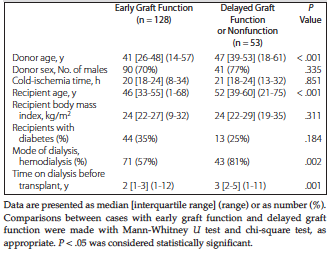
Table 1. Baseline Characteristics of the Deceased-Donor Kidney Grafts and Their Recipients (N = 181)
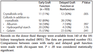
Table 2. Type of Fluids Used in Treating Donors
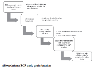
Figure 1. Flow Chart for the Inclusion of Cases in the Study
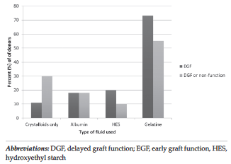
Figure 2. Diagram Showing the Outcomes of Early and Delayed Graft Function Depending on Which Fluids Were Used in Treating the Donor