Volume: 15 Issue: 1 February 2017 - Supplement - 1
FULL TEXT
Objectives: In this study, we report our experience at King Hussein Medical Center with Descemet stripping automated endothelial keratoplasty.
Materials and Methods: This study was conducted at King Hussein Medical Center of the Royal Medical Services during the period between January 2015 and January 2016. Patients who underwent Descemet stripping automated endothelial keratoplasty were enrolled in the study. We performed a comprehensive overview of the advantages of the surgery and analyzed the donor tissue selection criteria, indications, detailed surgical techniques of the procedure, and surgical outcomes.
Results: Our cohort included 13 patients. We had successful results for all of our patients, with final visual acuity of more than 6/12. The 2 indications for surgery were pseudophakic bullous keratopathy and Fuchs endothelial dystrophy.
Conclusions: Descemet stripping automated endothelial keratoplasty is becoming a revolutionary procedure in corneal surgery and replacing penetrating keratoplasty in many clinical situations.
Key words : Bullous, Endothelium, Fuchs, Recipient
Introduction
Partial thickness corneal surgery has evolved rapidly in the past 2 decades, replacing penetrating keratoplasty, the criterion standard for many decades.1 In 1998, posterior lamellar keratoplasty as first partial thickness procedure was used.2 Other partial thickness procedures are deep lamellar endothelial keratoplasty, Descemet stripping endothelial keratoplasty,3 Descemet stripping automated endothelial keratoplasty (DSAEK),4 Descemet membrane endothelial keratoplasty (DMEK),5 Descemet membrane automated endothelial keratoplasty,6 and DMEK without microtome.7
Descemet stripping automated endothelial keratoplasty is a modified technique of Descemet stripping endothelial keratoplasty, where a microkeratome is used for the donor dissection.8 It was first described in 2006 by Dr. Mark Gorovoy.4 In the procedure, Descemet membrane and endothelium are stripped from the host cornea and replaced with a donor button consisting of posterior stroma, Descemet membrane, and endothelium. Descemet membrane endothelial keratoplasty is a technique where only Descemet membrane and endothelium are used to replace host tissue of Descemet membrane and endothelium without posterior stroma. A modification of DMEK was described in 2009 where a rim of stroma was left at the periphery of the donor tissue using microkeratome (Descemet membrane automated endothelial keratoplasty) and in 2010 manually without microkeratome.7
Among all partial thickness procedures, DSAEK is the most common type of endothelial keratoplasty performed worldwide.8 The aim of our study is to report our experience at King Hussein Medical Center with DSAEK.
Materials and Methods
This study was conducted at King Hussein Medical Center of the Royal Medical Services during the period between January 2015 and January 2016. Patients who underwent DSAEK were enrolled in the study. For our patients, we used precut tissue (prepared by the eye bank using a Moria microkeratome; Moria Surgical, Antony, France). The donor tissue selection criteria were graft thickness < 130 μm, cap size of 9 mm, cell count of 3000 cells, age difference between recipient and host < 10 years, death to preservation < 12 hours, and death to surgery ≤ 5 days.
The surgeries were done under local peribulbar anesthesia, followed by the use of Honan balloon (The Lebanon Corp., Lebanon, PA, USA). The surgery technique that we used was started by creating temporal scleral incision with 5.5 mm width. Recipient descematorhexis was performed with the aid of reverse Sinskey hook and an anterior chamber maintainer. This was followed by inserting DSAEK tissue using a Busin glide (Moria) and pull-through technique. The temporal incision was sutured using 10/0 nylon suture, air was inserted, and complete air fill was maintained for 15 minutes. The air bubble size was reduced, and the patient was kept in the supine position for 1 hour and examined by slit lamp. Dilating drops (10% phenylephrine, 1% tropicamide) were used to avoid pupillary block. The patients were asked to stay in the supine position as much as possible during the first 24 hours to avoid graft detachment.
Patients were followed up postoperatively after1 day, 1 week, 1 month, and then monthly after that. They were prescribed local prednisolone ophthalmic and ofloxacin ophthalmic eye drops after surgery. Investigations done included pentacam and pachymetry.
Results
Thirteen patients were included in our series. Twelve of the patients had pseudophakic bullous keratopathy, and the remaining patient was phakic with Fuchs endothelial dystrophy. We had successful results for all of our patients with final visual acuity of more than 6/12.
Figure 1 shows the preoperative appearance of a patient with pseudophakic bullous keratopathy, and Figures 2 and 3 show the same patient’s postoperative appearance 1 day and 1 month after surgery. The patient’s final visual acuity was 6/9. The patient in our series who had Fuchs endothelial dystrophy showed corneal edema in right eye and guttata in left eye. Figures 4 and 5 show this patient’s pre- and postoperative appearances.
Discussion
Penetrating keratoplasty is a full-thickness corneal transplant procedure. It is still considered as the criterion standard transplant surgery by many surgeons worldwide. There are many disadvantages for the procedure, including expulsive choroidal hemorrhage (0.5%-1%), endothelial graft rejection and failure, decreased survival of regrafts, astigmatism, unexpected refractive outcomes, ocular surface-related problems, frequent visits, wound slippage and traumatic dehiscence, infection, and suture-related problems.
Many of the corneal pathologies are localized either anteriorly or posteriorly. Examples of anterior pathologies are keratoconus, viral keratitis, corneal stromal dystrophies, and degenerations. In such cases, a partial graft or deep anterior lamellar keratoplasty may be used for better visual outcome. Other indications for deep anterior lamellar keratoplasty are descemetocele, small perforations, and severe thinning where a tectonic graft could be used. Posterior pathologies where DSAEK are indicated include Fuchs endothelial dystrophy, pseudophakic and aphakic corneal edema, endothelial decompensation, failed grafts, iridocorneal endothelial syndrome, and posterior polymorphous corneal dystrophy.
Advantages of DSAEK include that it allows vision improvement to 6/9 to 6/12, has a lower rejection rate than penetrating keratoplasty, results in faster visual rehabilitation, uses a small incision and astigmatic neutral surgery, and maintains globe integrity and less wound dehiscence. However, DSAEK may still limit the best-corrected vision due to the donor lamellar interface.9-11
Figures 1 to 3 show our results for a patient with pseudophakic bullous keratopathy. The patient shown in Figures 4 and 5 was a 50-year-old male who had been referred with visual acuity of counting fingers of 0.5 m of the right eye and 6/6 of the left eye. Central corneal thicknesses measured 800 μm in the right eye and 690 μm in the left eye. He was phakic and had diffuse edema in his right cornea and guttata on the left. Pentacam showed anterior chamber depth (ACD) of 3.05 mm in the right eye. He underwent DSAEK, and his final visual outcome was 6/12 after 8 months.
Descemet stripping automated endothelial keratoplasty seems to be superior to penetrating keratoplasty in treating Fuchs endothelial keratoplasty, although primary graft failure may be more common. Visual recovery is faster, and major ametropia and astigmatism are not induced.10,12-14
The problem in phakic DASEK is the development in cataracts in such patients. In our opinion, this procedure should be done by expert surgeons. Usually patients who have phakic DSAEK often need phacoemulsification surgery within 18 to 24 months. Anterior chamber depth measurement is mandatory in such surgeries. Our recommendations are as follows: if ACD is > 3.0 mm, conduct DSAEK alone, depending on the refractive status of the patient; if ACD is 2.5 to 2.99 mm, perform either option, depending on the surgeon’s experience; and if ACD is 2.0 to 2.49 mm, perform phacoemulsification/DSAEK.
In conclusion, DSAEK is becom ing a revolutionary procedure in corneal surgery and is replacing penetrating keratoplasty in many clinical situations.
References:
- Cursiefen C, Kruse FE. Descemet's stripping with automated endothelial keratoplasty (DSAEK). Ophthalmologe. 2008;105(2):183-190.
CrossRef - PubMed - Melles GR, Eggink FA, Lander F, et al. A surgical technique for posterior lamellar keratoplasty. Cornea. 1998;17(6):618-626.
CrossRef - PubMed - Melles GR, Wijdh RH, Nieuwendaal CP. A technique to excise the Descemet membrane from a recipient cornea (descemetorhexis). Cornea. 2004;23(3):286-288.
CrossRef - PubMed - Gorovoy MS. Descemet-stripping automated endothelial keratoplasty. Cornea. 2006;25(8):886-889.
CrossRef - PubMed - Melles GR, Ong TS, Ververs B, van der Wees J. Descemet membrane endothelial keratoplasty (DMEK). Cornea. 2006;25(8):987-990.
CrossRef - PubMed - Price MO, Giebel AW, Fairchild KM, Price FW. Descemet's membrane endothelial keratoplasty: prospective multicenter study of visual and refractive outcomes and endothelial survival. Ophthalmology. 2009;116(12):2361-2368.
CrossRef - PubMed - Studeny P, Farkas A, Vokrojova M, Liskova P, Jirsova K. Descemet membrane endothelial keratoplasty with a stromal rim (DMEK-S). Br J Ophthalmol. 2010; 94(7): 909-914.
CrossRef - PubMed - Ang AY, Liu YC, Tan DT, Mehta JS. Descemet stripping automated endothelial keratoplasty with the EndoGlide Ultrathin graft insertion device. Expert Rev Med Devices. 2014;11(6):573-579.
CrossRef - PubMed - Price MO, Price FW. Descemet's stripping endothelial keratoplasty. Curr Opin Ophthalmol. 2007;18(4):290-294.
CrossRef - PubMed - Mau K. What DSAEK is going on? An alternative to penetrating keratoplasty for endothelial dysfunction. Optometry. 2009;80(9):513-523.
CrossRef - PubMed - Maier P, Reinhard T, Cursiefen C. Descemet stripping endothelial keratoplasty--rapid recovery of visual acuity. Dtsch Arztebl Int. 2013;110(21):365-371.
PubMed - Hjortdal J, Ehlers N. Descemet's stripping automated endothelial keratoplasty and penetrating keratoplasty for Fuchs' endothelial dystrophy. Acta Ophthalmol. 2009;87(3):310-314.
CrossRef - PubMed - Vedana G, Villarreal G Jr, Jun AS. Fuchs endothelial corneal dystrophy: current perspectives. Clin Ophthalmol. 2016;10:321-330.
PubMed - Zhang J, Patel DV. The pathophysiology of Fuchs' endothelial dystrophy--a review of molecular and cellular insights. Exp Eye Res. 2015;130:97-105.
CrossRef - PubMed

Volume : 15
Issue : 1
Pages : 124 - 127
DOI : 10.6002/ect.mesot2016.A208
From King Hussein Medical Center, Amman, Jordan
Acknowledgements: The authors declare that they have no sources of funding for
this study, and they have no conflicts of interest to declare.
Corresponding author: Amal Al-Thawabi, King Hussein Medical Center, Amman, PO
Box 393 Postal code 19152, Jordan
Phone: +962 797 835 222
E-mail: aalthawabi@hotmail.com.
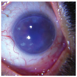
Figure 1. Preoperative Appearance of Patient with Pseudophakic Bullous Keratopathy
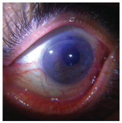
Figure 2. Patient With Pseudophakic Bullous Keratopathy Day 1 After Surgery
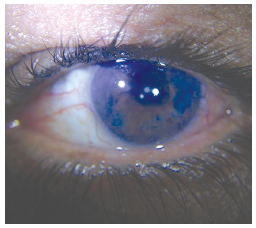
Figure 3. Patient With Pseudophakic Bullous Keratopathy Month 1 After Surgery
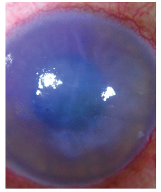
Figure 4. Patient With Fuchs Endothelial Dystrophy and Corneal Edema and Guttata Preoperatively
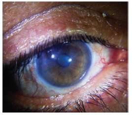
Figure 5. Patient With Fuchs Endothelial Dystrophy and Corneal Edema and Guttata Postoperatively