Volume: 15 Issue: 1 February 2017 - Supplement - 1
FULL TEXT
A 47-year-old male patient underwent living-related renal transplant. On day 3 posttransplant, without evidence of associated clinical symptoms, the patient’s serum creatinine levels had increased. The patient was given immunosuppressive medication, and a follow-up Doppler ultrasonography revealed hypoechoic areas in the inferior pole of the renal parenchyma. Eventually, on day 25, there was no perfusion in the superior and inferior poles of the transplanted kidney. No venous flow was shown in the middle segment, and only arterial vascularization with a high resistive index and negative diastolic phase was observed. Renal biopsy showed acute humoral rejection. This was interpreted as venous thrombosis secondary to acute humoral rejection. Tissue plasminogen activator infusion, plasmapheresis, and hemodialysis were administered. After 1.5 months, arterial flow returned to its normal pattern and the renal allograft recovered by gaining back its full vascularity at the end of month 8.
Key words : Acute humoral rejection, Renal transplant, Venous perfusion
Introduction
Kidney transplant is accepted as the final treatment for end-stage renal disease. However, it has many complications, including renal infarction, which is unique and seen during the early posttransplant period. Renal vein thrombosis is a critical and an uncommon complication that occurs in the first weeks after renal transplant. Renal infarction based on renal vein thrombosis has an incidence of 1%. Generally, mechanical causes underlie this complication, and it seldom derives from other causes, such as early severe rejection and hemostatic problems. No exact treatment is defined for this condition, and as a result the transplanted kidney is usually removed, unless prompt intervention is provided.1,2 The most common causes are mechanical, including the compression of the renal vein by collections such as hematoma or lymphocele and disturbances of the renal vein from stricture of the venous anastomosis or kinking of the vessel.3 The other causes consist of early severe rejection, hemostatic problems, or extension of ipsilateral iliac vein thrombosis.4
Renal vein thrombosis presents with nonspecific clinical and laboratory symptoms, such as lower quadrant pain, sudden hematuria, oliguria, hypotension, drop in hemoglobin, and elevation of serum creatinine.2 On the other hand, Doppler ultrasonographic evaluation can show diminished global perfusion and whether there is discontinued venous flow accompanied by an arterial pattern of high resistive index values and reversed diastolic phase. However, this Doppler ultrasonographic pattern is not characteristic of renal vein thrombosis, which can be observed during acute vascular rejection. It is hard to distinguish between these conditions by clinical and imaging methods during the early postoperative period.5
Case Report
We present a case of a 47-year-old male patient who underwent living-related renal transplant. The graft was from his wife, and it was implanted by anastomosing the renal vein of the graft to the left external iliac vein. This was his first and only transplant procedure.
On day 3 posttransplant, he showed an increase in serum creatinine levels (1.2 mg/dL). The patient presented with no other associated clinical symptoms. Doppler ultrasonography, by using Siemens Sonoline Antares (Siemens, Munich, Germany), performed the same day demonstrated vascular patency; however, renal pelvic mucosal edema was noted. Methylprednisolone (250 mg twice per day) was added to his immunosuppressive therapy. On day 7 posttransplant, a renal biopsy was performed due to high serum creatinine levels that could not be reduced. The biopsy revealed type 2a acute cellular rejection (Figure 1A). Antithymocyte globulin (140 mg) and cytomegalovirus prophylaxis were started for the treatment of acute rejection episode. Cytomegalovirus DNA polymerase chain reaction and urine culture results were negative. Follow-up Doppler ultrasonography revealed hypoechoic areas in the renal parenchyma of the inferior pole.
On day 25, the patient demonstrated no perfusion in the superior and inferior poles of the renal transplant and only the middle segment was vascularized with an arterial supply of high resistive index and negative diastolic phase (Figure 2). There was no venous flow in the middle segment, and this was interpreted as venous thrombosis secondary to acute humoral rejection. A second renal biopsy was performed, which later caused perirenal hematoma, resulting in acute humoral rejection (Figure 1B and 1C). Renal angiography was conducted the same day, and the main renal venous structures were found patent. Tissue plasminogen activator infusion was administered for possible thrombosis in the distal branches. In addition, the patient received plasmapheresis and hemodialysis.
On day 26, Doppler ultrasonography revealed minimal venous flow in the middle segment, showing a velocity of 26 cm/s. Arterial flow continued to show high resistive index, there was increased parenchymal echogenicity (grade 1/2), and parenchymal necrosis in the upper and lower poles of the transplanted kidney remained the same (Figure 3A). After 1.5 months, arterial flow returned to its normal pattern and the necrotic tissues showed significant atrophy; however, the rest of the kidney had hypertrophied and had returned to its normal size and configuration. At month 8, normal flow was observed (Figure 3B). The patient is alive with a functioning graft for more than 20 months.
Discussion
Renal vein thrombosis is an important and unique complication of renal transplant with an incidence of 1%, which usually ends in graft loss. It is defined more commonly with deceased-donor grafts (2%) than with living-related donor grafts (0.6%). It is also more common in female (1.75%) and adult recipients (1.2%) that in male (0.5%) and pediatric recipients (0%).2 Because it is an early postoperative complication that occurs in the first several weeks after transplant, there is an overlap of diagnosis between renal vein thrombosis and other similar clinically presented early postoperative complications, such as acute tubular necrosis or acute vascular rejection. Without prompt diagnosis and intervention, salvage possibility is poor, which may lead to renal allograft rupture and eventually to graft loss.
Renal vein thrombosis is a critical outcome of transplant that needs immediate surgical intervention.6 Therefore, in the early period posttransplant, together with clinical symptoms such as hematuria, oliguria, and hypotension, Doppler ultrasonographic findings of high resistance arterial flow with poor parenchymal perfusion can be used to consider renal vein thrombosis as an underlying reason of symptoms, and treatment should be done accordingly. Our patient, who presented with renal vein thrombosis due to acute humoral rejection, is unique regarding his graft gaining back its perfusion ability after a long interval of parenchymal necrosis, with restoration of graft to full function and size.
References:
- Gruber SA, Chavers B, Payne WD, et al. Allograft renal vascular thrombosis--lack of increase with cyclosporine immunosuppression. Transplantation. 1989;47(3):475-478.
CrossRef - PubMed - Fathi T, Samhan M, Gawish A, Donia F, Al-Mousawi M. Renal allograft venous thrombosis is salvageable. Transplant Proc. 2007;39(4):1120-1121.
CrossRef - PubMed - Koester BH, Köveker GB, Pötsch B, Becker HD. Hereditary thrombophilia as a cause of recurrent transplant thromboses. Chirurg. 1993;64(10):809-812.
PubMed - Schwieger J, Reiss R, Cohen JL, Adler L, Makoff D. Acute renal allograft dysfunction in the setting of deep venous thrombosis: a case of successful urokinase thrombolysis and a review of the literature. Am J Kidney Dis. 1993;22(2):345-350.
CrossRef - PubMed - Claudon M, Cosgrove D, Albrecht T, et al. Guidelines and good clinical practice recommendations for contrast enhanced ultrasound (CEUS) - update 2008. Ultraschall Med. 2008;29(1):28-44.
CrossRef - PubMed - Grzelak P, Kurnatowska I, Sapieha M, et al. Disturbances of kidney graft perfusion as indicators of acute renal vein thrombosis in contrast-enhanced ultrasonography. Transplant Proc. 2011;43(8):3018-3020.
CrossRef - PubMed

Volume : 15
Issue : 1
Pages : 244 - 246
DOI : 10.6002/ect.mesot2016.P117
From the 1Department of Radiology, the 2Department of Pathology, and the 3Department of General Surgery, Başkent University Medical Faculty, Ankara, Turkey
Acknowledgements: The authors declare that they have no sources of funding for this study, and they have no conflicts of interest to declare.
Corresponding author: Funda Ulu Ozturk, Başkent University Hospital, Department of Radiology, Mareşal Fevzi Çakmak St. No:45, Bahçelievler 06490, Ankara, Turkey
Phone: +90 555 223 6070
E-mail: nepandes@hotmail.com
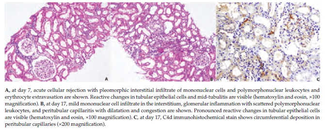
Figure 1. Day 7 and Day 17 Posttransplant Renal Biopsies
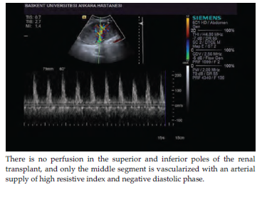
Figure 2. Day 25 Doppler Ultrasonographic Findings
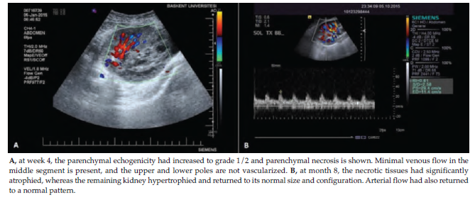
Figure 3. Week 4 and Month 8 Doppler Ultrasonographic Findings