Volume: 15 Issue: 1 February 2017 - Supplement - 1
FULL TEXT
Urinary tract infection is the most common complication after kidney transplant that can cause graft loss. An early diagnosis of urinary infections decreases morbidity and mortality. Besides clinical and laboratory examinations, ultrasonography is considered as the primary imaging modality for the diagnosis of urinary tract infections. Here, we report a 53-year-old woman who presented with fever and pain at surgical site. Ultrasonography examination showed multiple, ill-defined or irregularly margined hypoechoic areas within the cortex. Ultrasonography-guided percutaneous renal biopsy was performed. Histopathologic findings were compatible with acute tubulointerstitial nephritis and multiple abscesses. Ultrasonography is the most widely applied imaging modality for diagnoses of complications after renal transplant. Although ultrasonography findings of infections are generally nonspecific, it still plays an important role in the diagnosis of urinary infections after renal transplant.
Key words : Infectious complications, Kidney transplant, Urinary infections
Introduction
Infectious complications constitute the major cause of morbidity and mortality, especially during the postoperative period in patients undergoing solid-organ transplant. The prevalence of urinary tract infections is approximately 5% to 36% and may reach up to a 60% during the first year after transplant.1,2 Immunosuppressive medications are the major causes of infections among organ transplant recipients.3 Early diagnosis of urinary infections improves long-term graft survival. Ultrasonography is a relatively inexpensive and noninvasive modality that can be used to diagnose complications and to provide follow-up after transplant. Ultrasonography examination includes size and echogenicity of kidney, collecting system, and ureter, postoperative collections, and evaluation of vascular structure with color and spectral Doppler ultrasonography. Ultrasonography may also be used for guided renal biopsy and drainage of peritransplant collections. Here, we report ultrasonography findings in a patient with acute tubulointerstitial nephritis and multiple parenchymal abscesses after renal transplant.
Case Report
A 53-year-old woman, who had undergone kidney transplant 6 months earlier, presented with fatigue, fever, leukocytosis, and pain at surgical site. Because of recurrent urinary tract infections, right nephrectomy operation had been performed 10 days before presentation. No recent trauma and medical interventions had been reported. Klebsiella pneumoniae and extended-spectrum β-lactamase-producing Escherichia coli were found in the urine culture. Ultrasonography examination showed heterogeneous parenchyma with multiple, ill-defined or irregularly margined nonvascular hypoechoic areas within the cortex (Figure 1). No dilatation or mucosal thickening in the urine collecting system was noted. The vascular structure and flow quantification of transplanted kidney were normal in color and spectral Doppler ultrasonography examination. No perinephric fluid collection was detected. Ultrasonography-guided percutaneous renal biopsy was performed. Histopathologic findings were consistent with diffuse, severe, and acute tubulointerstitial nephritis and multiple abscesses.
Discussion
The treatment of choice for patients with end-stage renal disease is renal transplant. Urinary tract infection is a common complication after kidney transplant, often associated with graft loss and mortality.4 Some studies have shown more urinary tract infections in female patients than in male patients. In addition, older patients have a higher prevalence of urinary tract infections than younger patients. The most common cause of urinary tract infections is bacteria. Infections caused by fungi or viruses can also been seen, but less commonly.1
In addition to clinical and laboratory findings, imaging modalities, including ultrasonography, computerized tomography, and magnetic resonance, play important roles in diagnoses of urinary infections after renal transplant. Ultrasonography is considered as the primary imaging technique in the evaluation of kidney transplants. Ultrasonography is noninvasive, inexpensive, and relatively painless and can be easily applied at bedside. The relatively low cost and safety of ultrasonography allow serial examinations, which may be necessary during intraoperative or postoperative periods and for follow-up examinations. Ultrasonography may also be used as a guide for diagnostic and therapeutic interventions. A baseline ultrasonography evaluation is performed in the first 24 to 48 hours after transplant.5 The sonographic appearances of infections and abscesses are quite variable. Focal pyelonephritis may appear as hypoechoic or hyperechoic focal areas. These findings are nonspecific and can be misdiagnosed as infarction or rejection.3,4 A parenchymal or perinephric abscess complicates only a minority of cases. Abscesses may be seen as fluid-filled, complicated cysts at ultrasonography. Gas within the collection or hypoechoic focal areas in the parenchyma is highly suggestive for abscesses.3,4 Abscesses may be treated with either ultrasonography- or computed tomography-guided percutaneous drainage.5
Ultrasonography is the most widely applied imaging modality for diagnosis of complications and follow-up after kidney transplant and to guide treatment. Although sonographic findings of infections and abscesses are generally nonspecific or variable, ultrasonography still plays an important role in the diagnosis of urinary infections after renal transplant, due to advantages of being cost-effective and safe, having an easy application at bedside, and being available for serial examinations in intraoperative or postoperative periods.
References:
- Gondos AS, Al-Moyed KA, Al-Robasi AA, Al-Shamahy HA, Alyousefi NA. Urinary tract infection among renal transplant recipients in yemen. PloS One. 2015;10(12):e0144266.
CrossRef - PubMed - Espinar M, Miranda IM, Costa-de-Oliveira S, Rocha R, Rodrigues AG, Pina-Vaz C. Urinary tract infections in kidney transplant patients due to Escherichia coli and Klebsiella pneumoniae-producing extended-spectrum β-lactamases: risk factors and molecular epidemiology. PloS One. 2015;10(8):e0134737.
CrossRef - PubMed - Akbar SA, Jafri ZS, Amendola MA, Madrazo BL, Salem R, Bis KG. Complications of renal transplantation. Radiograph. 2005;25(5):1335-1356.
CrossRef - PubMed - Nixon JN, Biyyam DR, Stanescu L, Phillips GS, Finn LS, Parisi MT. Imaging of pediatric renal transplants and their complications: a pictorial review. Radiograph. 2013;33(5):1227-1251.
CrossRef - PubMed - Kolofousi C, Stefanidis K, Cokkinos DD, Karakitsos D, Antypa E, Piperopoulos P. Ultrasonographic features of kidney transplants and their complications: an imaging review. ISRN radiol. 2013;2013:480862.
CrossRef - PubMed

Volume : 15
Issue : 1
Pages : 247 - 248
DOI : 10.6002/ect.mesot2016.P118
From the 1Department of Radiology and the 2Department of General Surgery, Baskent University School of Medicine, Ankara, Turkey
Acknowledgements: The authors declare that they have no sources of funding for this study, and they have no conflicts of interest to declare.
Corresponding author: Sehnaz Tezcan, Baskent University Hospital, Department of Radiology, Maresal Fevzi Cakmak st. No:45, Bahcelievler, 06490, Ankara, Turkey
Phone: +90 533 455 55 33
E-mail: sehnazce@yahoo.com
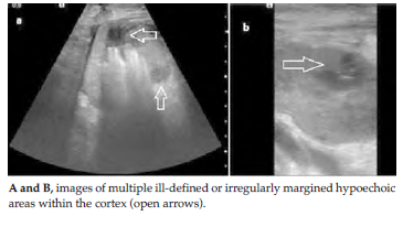
Figure 1. Ultrasonography Examination of Transplanted Kidney