Volume: 13 Issue: 3 November 2015 - Supplement - 3
FULL TEXT
Objectives: Assess the safety and accuracy of renal static scintigraphy with introduction of the amendment coefficient for decay of 99m metastable (Tc–99m) at the calculation formula of capture percent Technemec 99m.
Materials and Methods: This study analyzes retrospectively the results of renal static scanning of 44 patients. Of this group of patients, 21 are Chornobyl Nuclear Power Station accident liquidators who have arterial hypertension and chronic pyelonephritis. Renal scanning was carried out with Genesys EPIC single-photon emission computed tomography system. Calculation of the capture percent of the radiopharmaceutical Technemec 99m was executed in the standard way with introduction of the amendment coefficient Tc–99m to decay at the calculation formula of capture percentage Technemec.
Results: Significant differences by standard calcu-lation method took place in the both the Chornobyl Nuclear Power Station accident liquidators and persons who were not subjected to the influence of a small radiation dosage (P < .05).
Conclusions: A small radiation dose did not affect kidney function (P > .05) with introduction of the amendment coefficient for decay of technetium 99m; therefore, static scintigraphy is a safe method for assessing kidney structure and morphology. Introduction of amendment for decay of Tc–99m allowed false positive results in 11.9% of renal function assessments for patients with arterial hypertension and kidney dysfunction (P < .05). Calculation of the specific capture percent of radiopharmaceutical on 1 cm2 of visualization of renal parenchyma enabled a 29% increase in renal function identification accuracy.
Key words : Gamma camera imaging, Renal static scintigraphy
Introduction
Modern medical visualization methods are expensive and require strong and specific evidence for application. Technologies like magnetic resonance imaging (MRI) may also carry risks of its radiopharmaceutical (gadolinium) damaging a target organ such as a kidney.1 This has often led to false results when assessing pathological processes.
Nuclear medicine scans are less expensive and provide numerical information that often is unobtainable using other imaging techniques. The most sensitive test to define renal scar damage and kidney function is renal static scanning with Tc–99m–labeled dimercaptosuccinic acid (DMSA). In clinical practice, a Tc–99m DMSA scan is considered the standard reference method in the qualitative assessment of renal cortical lesions. However, more often it is used as a dynamic scan to assess kidney function.
Chronic pyelonephritis the outcome for continuing nonobstructive or obstructive pyelonephritis. Damaging processes lead to subsequent formation of kidney parenchymal scars. The degree of damage is even more expressed if urine becomes infected.2,3 Damage is closely interconnected with deformation of renal calices. Distortion and extension of renal calices occur due to damage of the kidney pyramids. As a rule, damage tends to ward the upper and lower kidney poles because these sites are more inclined to intrarenal reflux. Renal cortical and medullar layers became thinner at the damaged area, and the kidney becomes small and thin in its longitudinal and cross sizes.
Kidney damage can be defined with radiologic methods, such as ultrasound, intravenous urography, renal isotope scanning, or computed tomography (CT).These technologies currently comprise radio-logic semiotics for pyelonephritis.1-4
The renal static scintigraphy scan enables us to obtain morphologic data
about the organ (its localization, form, and size) to define its general
functional abilities. The radiopharmaceutical drugs arrive in the kidney via
afferent arterioles, and it is removed by proximal kidney tubules. Its
distribution is in a direct ratio to renal blood flow. One of the most
widespread radiopharmaceuticals used in renal scanning is Tc–99m DMSA. Its
standard calculation of capture percentage is as follows:
P1=[100 × (A-B)/C] × 1.73/S, where is P1 – a capture
percent of radiopharmaceutical by renal parenchyma; (A) impulse accounted from
renal area (impulses/min); (B) impulse accounted from background area
(impulses/min); (C) an activity difference at impulses/minute from a syringe and
after intravenous injection of radiopharmaceutical; 1.73 – a standard body
surface area; S – a patient body surface area determined with a growth-weight
nomogram for estimating body surface area.3,4
The optimal time to carry out a renal scan is 1 to 3 hours after intravenous injection of Tc–99m DMSA. The time duration for a renal scan is 5 to10 minutes. The process accumulation Tc–99m DMSA reflects renal function, regardless of specific physiological function. The average capture percent Tc–99m DMSA by an intact kidney is 3.90% ± 0.20%. For patients with essential hypertension, it is 1.78% ± 1.01%; patients with chronic pyelonephritis is decrease capture to 1.77% ± 0.29%. According to different data, the right kidney size is 10.9 ± 0.8 cm with a width that is 6.3 ± 0.5 cm; the left kidney size is 11.2 ± 0.9 cm with a width that is 6.4 ± 0.5 cm. The cross size of a kidney parenchyma is 4.4 ± 0.3 cm from the general kidney size.3-6 Calculation of the specific capture percent of radiopharmaceutical on 1 cm2 of visualization of renal parenchyma enables us to increase renal function identification accuracy by 29%.
The last major achievement in radionuclide diagnostics is application of single-photon emission computed tomography (SPECT). High-resolution imaging with pinhole collimators and SPECT is not possible with radiopharmaceuticals other than Tc–99m because of rapid transit times. The calculation of capture percentage of a radiopharmaceutical requires introduction of the amendment for organ depth because earlier results did not depend on the spatial arrangement of the organs. From our point of view, introduction of the amendment coefficient for the decay of Tc–99m is useful, and it enables us to carry out several additional impulse accounts at different positions. This requires additional time for scanning; however, it improves functional assessment in the case of kidney dystopia.
Radionuclide diagnostics of the urinary system are the most available, functional, and effective method for diagnosing renal pathological and general hemodynamic change in patients with arterial hypertension and chronic inflammation.5-7
Materials and Methods
In this study, we conducted cross-section analyses of the results of renal static scanning of 44 patients that had been observed in the Hospital for Persons with Disabilities from the Patriotic War (Table 1; Figures 1-2). Of this group of patients, 21 (ages, 35.8 ± 5.9 y) were liquidators at the Chornobyl Nuclear Power Station. They had hypertension (arterial pressure systolic 151.0 ± 21.1 mm Hg and diastolic 92.4 ± 13.7 mm Hg) and kidney disease (creatinine clearance 96.4 ± 15.6 mL/min; urea 5.4 ± 0.7 mmol/L). Average received radiation dosage exposure amounted to 0.10 ± 0.05 mSV.
Renal scanning was carried out with a Genesys EPIC single-photon emission computed tomography (SPECT) system (Philips/ADAC Labs, Milpitas, CA, USA) with Pegasys software (Open Source GNU General Public License https://bioinforatics.ubc.ca/ pegasys). The gamma camera is equipped with a low-energy high-resolution parallel collimator. For statistical scans, the matrix size was 286 × 286 × 26 cm in a power window of 140 KeV.
The renal static scan began with an intravenous injection the Technemec 99m (MEDI-RADIOPHARMA, Ltd., Hungary and DIAMED, Russia) at a dosage of 105 MBq for 70 kg of a patient weight. Two hours elapsed until the start of the investigation. Average radiation dose was 0.26 mSv. Technemec 99m consists of the Tc–99m complex with a mix of dimercaptosuccinic acid (1 mg) and 2-chloridetin (0.3 mg). For the most part, Tc–99m binds with serum proteins. The main way Tc–99m leaves the kidney is via tubular excretion and partially glomerular filtration as well. Renal excretion is slow. The radiopharmaceutical is prepared under aseptic conditions by adding 5 mL eluate from Tc–99m generator into the bottle with a reagent.
Calculation the percentage capture of Technemec Tc–99m radiopharmaceutical is executed in the stan-dard way and with introduction of the amendment coefficient for Tc–99m decay at calculation formula for the percentage capture of Technemec Tc–99m.
P1 = [100 × (A-B) × e0.693 × t/T)/ C] × 1.73/S, where P1 – a capture percent of the radiopharmaceutical by the kidney, taking into account amendment coefficient for decay of Tc–99m; (A) impulses accounted from renal area (impulses/min); (B) impulse accounted from background area (impulses/min); (C) an activity difference of impulses/minute from syringes until and after intravenous injection of radiopharmaceutical; 1.73 – standard body surface area; S – patient body surface area, which is determined with a growth-weight nomogram for estimating body surface area; e0.693 × t/T – amendment coefficient for decay of Tc-99m, where t – time from injection moment of Technemec Tc–99m until research start; T - half-life period of Tc–99m.
The study was approved by the Ethical Review Committee of the Medical Center. All of the protocols conformed to the ethical guidelines of the 1975 Helsinki Declaration. Written informed consent was obtained from all subjects.
Results
Standard accounting method for percentage capture of Tc–99m showed significant differences in accumulation of Tc–99m in renal parenchyma in patients who did not undergo small radiation dose (SRD) (Table 2). The proportion of patients who had been exposed to a SRD with low isotope accumulation is 0.83, and the patient arm who did not undergo a SRD is 0.62. Assessment of the differences between both proportions were defined as P = (-0.21); 95% CI is [-0.41; -0.01], by the Fisher exact test is Z = (-2.178; P < .05).
Proportion of patients with exposure to coefficient for decay of Tc–99m is 0.38; for patients not exposed to a SRD it is 0.52. Assessment of the differences between the proportions shows P = –0.14; 95% CI is [-0.336: 0.056], by the Fisher exact test is (Z = 1.324; P > .05).
Proportion of patients after a SRD exposure with 0.38 and without coefficient for decay of Tc–99m is 0.62. Assessment of difference between proportions has showed: P = .24 ± .20; 95% CI is [0.445: 0.034], by the Fisher exact test is (Z = 2.18; P < .05).
Proportions of patients who were not SRD-exposed with 0.52 and without coefficient for decay of Tc–99m is 0.83. Assessment of difference between proportions has showed: P = .30 ± 0.18; 95% CI is [0.48:0.12], by the Fisher exact test is (Z = 3.113; P < .05).
Discussion
In view of the high quantity of false negative results, DMSA kidney scanning should follow each negative renal ultrasound examination. Numeric information about kidney function is obtained with DMSA, and this is unique. There are powerful arguments for carrying out renal scanning in each case of infection in upper and lower urinary tracts. This is especially important in the case of recurrent infection in the urinary tract owing to repeated infection by the same or other infection agents or bacterial persistence by the same infection agents from within urinary tracts.1-3
Renal static scintigraphy is advised for assessment of the damaged mass (eg, cyst, tumor, or pseudocyst), for differential diagnostics of renal sinus cysts such as the parapelvical or peripelvical cysts from hydronephrosis, for congenital disease (eg, kidneys are fused together or ectopic), for an assessment of renal function at the damaged region or part of each kidney, for definition of a kidney scarring (eg, an infection consequence).2,4,6,7
The radiologic semiotics of the left and right kidneys are influenced by chronic inflammatory processes in the same patient and can be different; therefore, in this review, we estimated each kidney separately. According to the chronic pyelonephritis classification, we tried to create radionuclear semiotics concerning stages of the disease. According to this classification, Chornobyl accident liquidators and the persons not subjected to the influence of a SRD, but have symptomatic arterial hypertension in combination with chronic pyelonephritis, have different stages of pathological change.
In conclusion, static scintigraphy is a safe method for assessing kidney function. A small radiation dose did not affect kidney function (P > .05) with introduction of the amendment coefficient for decay of technetium 99m. Introduction of amendment for decay of Tc-99m allowed false positive results on 11.9% of the cases to assess of renal function of patients with arterial hypertension and kidney dysfunction (P < .05).
References:
- Chemecky, CC, Berger BJ. Laboratory Tests and Diagnostic Procedures. 5th ed. St. Louis, MO: Saunders; 2008.
- Reynard J, Brewster S, Biers S. Oxford Handbook of Urology. Practical Guidance for Diagnostics and Management. New York, NY: Oxford University Press; 2007.
- Tsvelev YV, Petrova SB. Urological Gynecology: Practical Guidance. Sankt Petersburg, Russia: Foliant Publishing House; 2006:272.
- Shihi G. Nuclear Medicine. Under edition Shlygina OE, Borisenko, AR. “Sansam” Publishing House; Almaty, 2006:136. (Part I) and 2008:288. (Part II).
- Resnick MI, Novick AC. Urology Secrets. Nevsk Dialect Publishing House; Russia, Sankt-Petersburg, 2000:352.
- Resnick MI, Sheffer AJ. Urology, 68 Cases from Practice. Nevsk Dialect Publishing House; Russia, Sankt-Petersburg, 2002:293.
- Shayman, JA. Renal Pathophysiology. BINOM Publishing House and Nevsk Dialect Publishing House; Russia, Sankt-Petersburg, 2001:206.

Volume : 13
Issue : 3
Pages : 69 - 73
DOI : 10.6002/ect.tdtd2015.P19
From the Department of Science Management, National Research Medical Center,
Astana, Kazakhstan
Acknowledgements: The author has no conflicts of interest to declare. No
funding was received for this study.
Corresponding author: Gulziya Ismailova, MD, PhD, Department of Science
Management, National Research Medical Center Avenue Abylay-khan 42, 010009,
Astana, Kazakhstan
Phone: +77 172 577 647; +77 018 228 188
Fax: +77 172 232 927
E-mail: dr.ismailova@mail.ru
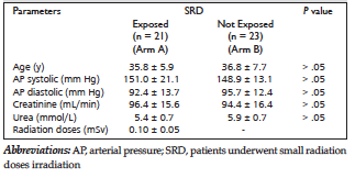
Table 1. General Characteristics of Patients
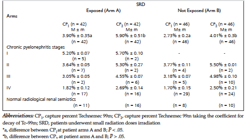
Table 2. Capture Percent Technemec 99m Relative to Chronic Pyelonephritis Stages
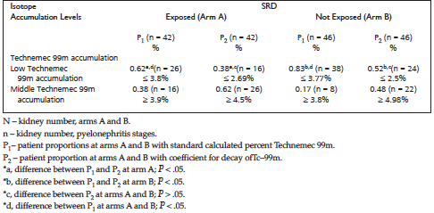
Table 3. Observed Patient Proportions Relative to Capture Percent Technemec 99m
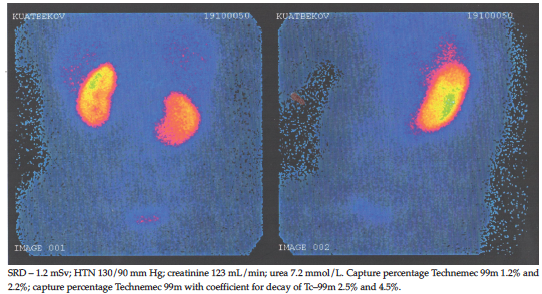
Figure 1. Static Scintigraphy of Patient Liquidator at the Chornobyl Nuclear Power Station (1988)
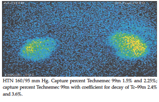
Figure 2. Static Scintigraphy of Patient