Volume: 13 Issue: 3 November 2015 - Supplement - 3
FULL TEXT
Objectives: Computed tomography is required for selection of living donors for kidney transplant. We assessed the diagnostic relevance and effectiveness of multiphase contrast-enhanced computed tomography angiography for evaluating patients before and after transplant.
Materials and Methods: Thirty-two potentially living kidney donors (15 men and 17 women) underwent multiphase computed tomography angiography for evaluation before kidney transplant and 2 posttransplant recipients underwent this test owing to abnormal vascularization. Computed tomography angiography was used to determine parenchymal blood-flow conditions and vascular architecture of the kidney grafts. All of the 34 patients underwent prestudy Doppler ultrasonography.
Results: Additional renal arteries were found in 11 patients (32.3%). From them, a single additional renal artery, situated from the aorta to the lower segment of the kidney, was observed in 8 (72.7%) patients; dual additional renal arteries with equivalent caliber trunks were observed in 3 (27.3%) patients. An additional renal artery occurred more often in women in 6 (54.5%) donors. Concomitant renal pathology was detected in 3 (9.3%) of 32 donors: simple cysts in 2 donors and hydronephrosis transformation in 1 donor. These 3 cases represented an accidental discovery and were therefore excluded from the list of donors. In addition, all donors underwent excretory-phase computed tomography, and their renal function and urinary collecting system results were evaluated. Two recipients with graft dysfunction presented with decreased venous return and delayed arterial filling of the renal arteries that manifested as delayed contrast in the kidney graft medulla. Renal parenchymal ischemic zones were consistent with microvasculature thrombosis, the most common cause of vascular complications in these recipients.
Conclusions: Multiphase computed tomography angiography is a necessary tool for assessing the structure and condition of vascular architecture in kidney donors and recipients. Individuals with ambiguous Doppler results, this test should be required; it is also warranted when renal graft vascular complications are suspected.
Key words : Graft dysfunction, Multiphase computed tomography angiography, Renal transplant
Introduction
Computed tomography (CT) is a mandatory method of investigation for selecting donors for kidney transplant.1 It is also valuable for visualizing main and segmental renal blood flow (arterial and venous) with multiphase contrast-enhanced CT angiography (MP-CTA) to define the approach to further treatment. We assessed the diagnostic relevance and effectiveness of MP-CTA in the evaluation of patients before and after kidney transplant.2
Materials and Methods
The study was approved by the Ethical Review Committee of the Institute. All of the protocols conformed to the ethical guidelines of the 1975 Helsinki Declaration. This was a retrospective analysis all kidney transplants conducted in our hospital during 2012 to 2014. Thirty-two living kidney donors (15 men and 17 women) underwent MP-CTA before kidney transplant for pretransplant evaluation, and 2 transplant recipients (both men) underwent MP-CTA after transplant owing to abnormal graft vascularization. All of the 34 participants had undergone prestudy Doppler ultrasonography. Parenchymal blood-flow conditions and vascular architecture of the kidney grafts were determined using MP-CTA. Multiphase contrast-enhanced CT angiography studies were conducted on a 64-slice Aquilion64 CT scanner (Toshiba, Japan) in conjunction with a digital processing system, with results processed on the workstation using the VitreaEnterprise Suite software package (Vital Images, Minneapolis, MN, USA). Analyses of images included a multiplanar reconstruction of the defect in various projections. All patients were injected with Ultravist370 (Bayer: Pharma AG, 13342, Berlin, Germany) contrast media (iodine 370 mg dosed at 1.5 mL/kg).
Results
In our study, an additional renal artery or arteries were found in 11 donors (32.3%). A single additional renal artery, situated from the aorta to the lower segment of the kidney, was observed in 8 donors (72.7%). Dual additional renal arteries, with equivalent caliber trunks, were observed in 3 donors (27.3%). An additional renal artery was more common in women (n = 6 [54.5%]). Concomitant renal pathology was detected in 9.3% of donors: simple cysts in 2 donors and hydronephrosis transformation in 1 donor. This latter case was an accidental discovery and therefore was excluded from the number of donors. In addition, all patients underwent excretory-phase CT and were evaluated for renal function and urinary collecting system.
Multiphase contrast-enhanced CT angiography was conducted if graft dysfunction or vascular complications were suspected. Patients who underwent MP-CTA showed a decrease in venous return with timely arterial filling of the renal arteries, manifesting as delayed contrast in the kidney graft medulla. Renal parenchymal ischemic zones were consistent with microvasculature thrombosis, which was the most common cause of vascular complications in our study population.
In 1 case, a patient who received a kidney from a related living-donor experienced delayed graft function owing to acute kidney injury. Doppler ultrasonography showed lack of vascular blood flow in the graft, but a fuller picture of renal graft blood flow was seen on MP-CTA, which determined the ischemic upper pole of the kidney graft (Figure 1). During all phases of MP-CTA, uneven contrast in the upper pole of the kidney graft was observed as a local area (2.5 × 2.0 cm) of reduced density. Notable low-intensity contrast was seen in the lumen of the artery and vein of the kidney graft and no image showed renal artery stenosis or thrombosis. These findings were regarded as ischemic areas with necrotic foci, which confirmed the clinical diagnosis of acute tubular necrosis.
Discussion
To visualize the number of renal arteries and veins, as well as the nature of their division, CT angiography is the least invasive and most informative imaging method.3 The advantage of CT angiography is maximum possible resolution in a 3-dimensional image, which enables detection of minimal changes in internal organs, including blood vessels.4 In this study, the status of main and segmental blood flow in the kidney graft was determined using CT angiography, whereas Doppler ultrasonography did not adequately depict blood flow. The visualization of blood flow in the kidney graft using MP-CTA angiography allows graft rejection and further surgery to perhaps be avoided.5 In addition, in screening potential kidney donors, MP-CTA could identify comorbidities that were used as exclusion criteria.6
Conclusions
Multiphase contrast-enhanced CT angiography is a necessary tool for estimating the structure and condition of the vascular architecture of the kidney in donors and recipients. In cases where Doppler ultrasonography results are ambiguous, MP-CTA is warranted. Multiphase contrast-enhanced CT angiography also should be carried out when renal graft vascular complications are suspected.
References:
- Napoli A, Fleischmann D, Chan FP, et al. Computed tomography
angiography: state-of-the-art imaging using multidetector-row technology. J
Comput Assist Tomogr. 2004;28(suppl 1):S32-S45.
CrossRef - PubMed - Helck A, Bamberg F, Sommer WH, et al. Optimized contrast volume for
dynamic CT angiography in renal transplant patients using a multiphase CT
protocol. Eur J Radiol. 2011;80(3):692-698.
CrossRef - PubMed - Patil UD, Ragavan A, Nadaraj, et al. Helical CT angiography in
evaluation of live kidney donors. Nephrol Dial Transplant.
2001;16(9):1900-1904.
CrossRef - PubMed - Frauenfelder T, Wildermuth S, Marincek B, Boehm T. Nontraumatic emergent
abdominal vascular conditions: advantages of multi-detector row CT and
three-dimensional imaging. Radiographics. 2004;24(2):481-496.
CrossRef - PubMed - Singh AK, Sahani DV. Imaging of the renal donor and transplant
recipient. Radiol Clin North Am. 2008;46(1):79-93, vi.
CrossRef - PubMed - Pham PC, Wilkinson AH, Pham PT. Evaluation of the potential living
kidney donor. Am J Kidney Dis. 2007;50(6):1043-1051.
CrossRef - PubMed

Volume : 13
Issue : 3
Pages : 88 - 90
DOI : 10.6002/ect.tdtd2015.P48
From the 1Department of Radiology, JSC National Research Medical
Center, Astana, Kazakhstan; and the 2Department of Extracorporeal
Hemocorrection, JSC National Scientific Medical Research Center, Astana, 010009,
Kazakhstan
Acknowledgements: The authors declare that they have no sources of
funding for this study, and they have no conflicts of interest to declare.
Corresponding author: Timur Sarsengaliyev, Department of Radiology, JSC
National Scientific Medical Research Center, 010009, Ave Abylay-khan #42,
Astana, Kazakhstan
Phone: +77 172 577 481
Fax: +77 172 232 927
E-mail:
timur_sarsengaliev@mail.ru
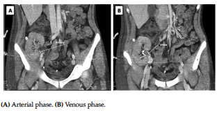
Figure 1. Multiphase Contrast-Enhanced Computed Tomography Angio - graphy of a Kidney in Recipient After Transplant